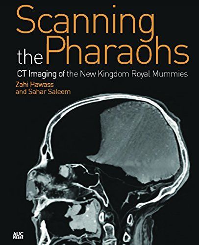
Scanning the Pharaohs CT Imaging of the New Kingdom Royal Mummies
The royal mummies in the Cairo Museum are an important source of information about the lives of the ancient Egyptians. The remains of these pharaohs and queens can inform us about their age at death and medical conditions from which they may have suffered, as well as the mummification process and objects placed within the wrappings. Using the latest technology, including Multi-Detector Computed Tomography and DNA analysis, co-authors Zahi Hawass and Sahar Saleem present the results of the examination of royal mummies of the Eighteenth to Twentieth Dynasties. New imaging techniques not only reveal a wealth of information about each mummy, but render amazingly lifelike and detailed images of the remains. In addition, utilizing 3D images, the anatomy of each face has been discerned for a more accurate interpretation of a mummy's facial features. This latest research has uncovered some surprising results about the genealogy of, and familial relationships between, these ancient individuals, as well as some unexpected medical finds. Historical information is provided to place the royal mummies in context, and the book with its many illustrations will appeal to Egyptologists, paleopathologists, and non-specialists alike, as the authors seek to uncover the secrets of these most fascinating members of the New Kingdom royal families.